Introduction
The skin is the most superficial part of the body. The signs of ageing are most visible in the skin. Although, ageing skin is not a threat to a person, it can have a detrimental effect on the psychology of a person. A look into the causes of skin ageing, the available treatments and preventive measures for this inevitable change is important to help both the already aged, as well as, the youth.
This is a 4 part article in which:
- Part 1 – Discusses the structure of skin and its different components
- Part 2 – Discusses cutaneous ageing and its causes
- Part 3 – Discusses the characteristics of ageing skin and the changes in skin appearance (current article)
- Part 4 – Discusses products and treatments for skin ageing
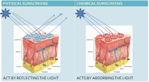
a) Sunscreen Agents
b) Moisturizers
c) Antioxidants
d) Make Up
e) Dermal Fillers
f) Chemical Peels
g) Botulinum Toxin
h) Estrogen and Hormonal Treatments
i) Plastic Surgery
Part 3 – Characteristics of the Ageing Skin
The changes undergone by skin as it ages, occurs throughout the epidermis, dermis and the subcutaneous tissue.

Epidermal Changes
The intersection of epidermis and dermis is known as the Dermal Epidermal Junction (DEJ). The DEJ is known to be altered with age. Aged epidermis shows a flattened DEJ with a correspondingly diminished connective surface area (2.64 mm2 to 1.9 mm2). Loss of DEJ surface area may contribute to increased fragility of the skin and may also lead to reduced nutrient transfer between dermal and the epidermal layers.
According to studies, on sun exposed and protected skin, from volunteers aged 6‐ 84 years, it revealed that the epidermal thickness to be constant across the decades, but with thickness found to be greater in sun exposed skin. Spinous layer of a wrinkle was seen to be thinner at the base than flanks. The epidermis also showed fewer keratohyaline granules at the base as compared to flanks.
Decreased cell turnover‐ the epidermal turnover rate slows from 30% from 50%. The stratum corneum transit time was shown to be 20 days in young adults, and 30 or more days in older adults. Such a cell cycle lengthening in older adults coincides with a protracted stratum corneum replacement time, epidermal atrophy, slower wound healing, and often less effective desquamation. Older patients would thus require double the time to reepithelialize following dermabrasion resurfacing procedures compared to younger patients. The cascade of changes related to decelerated cell turnover results in development in heaps of corneocytes that render a rough and dull skin surface.
Dermal Changes
Approximately 20% of dermis thickness disappears in older adults. Normal aged dermis is characterized by changes in collagen production and development of fragmented elastic fibers. A photoaged dermis exhibits disorganized collagen fibrils and accumulation of abnormal elastin containing material.
COLLAGEN
The primary structural component of the dermis and the most abundant protein found in humans, collagen is responsible for conferring strength and support to human skin. Over time, the structural proteins and main components of the skin deteriorate, resulting in the cutaneous signs of ageing. Intrinsically aged skin is characterized by epidermal and dermal atrophy as well as flattening of the rete ridges. It is well known, however, that alterations in collagen play an integral role in the ageing process. This, in turn, partly explains the popularity of collagen‐containing products intended for ‘anti‐ageing’ purposes.
Of the dry skin mass, 70% is comprised of collagen. In aged skin, collagen is characterized by thickened fibrils, organized in rope‐like bundles, that appear to be in disarray in comparison to the pattern observed in younger skin. In addition, lower levels of collagen are synthesized, in vivo and in vitro, by aged fibroblasts. The ratio of collagen types found in human skin also changes with age. In young skin, collagen I comprises 80% and collagen III comprises about 15% of total skin collagen; in older skin, the ratio of Type III to Type I collagen has been shown to increase, due, significantly, to an appreciable loss of collagen I. In addition, the overall collagen content per unit area of skin surface is known to decline approximately 1%/year.
In irradiated skin, collagen I levels have been shown to be reduced by 59%; this reduction was found to be linked to the extent of photodamage. Although collagen I is the most abundant and significant collagen type found in the skin, the effects of ageing are seen in other types of collagen in human dermis. An integral constituent of the DEJ, collagen IV imparts a structural framework for other molecules and plays a key role in maintaining mechanical stability. No significant differences have been found in collagen IV levels in sun‐exposed skin compared to unexposed skin, but significantly lower levels of collagen IV have been identified at the base of wrinkles in comparison to the flanks of the same wrinkles.
The mechanical stability of the DEJ may be adversely affected by this loss of collagen IV, thereby contributing to wrinkle formation. Collagen VII is the primary constituent in anchoring fibrils that attach the basement membrane zone to the underlying papillary dermis. In one study, a significantly lower number of anchoring fibrils were identified in patients with chronically sun‐exposed skin in comparison to normal controls. It was theorized by the researchers that wrinkles may form as a result of a weakened bond between the dermis and epidermis, due to anchoring fibril degradation. A more recent study showed such a loss of collagen VII to be more marked in the base of the wrinkle (as seen with collagen IV in the same study).
In the last 15 years, the pathogenesis of UVR induced collagen damage has been well understood and characterized. For instance, it is known that UVR exposure significantly up‐regulates the synthesis of several types of collagen‐degrading enzymes known as matrix metalloproteinases (MMPs). First, UV exposure leads to an increase in the amount of the transcription factor c‐jun; cfos, the other transcription factor involved in this mechanistic chain, is already abundant without UV exposure. Activator protein‐1 (AP‐1) is then formed by the combination of c‐jun and cfos. In turn, AP‐1 activates the MMP genes, which stimulate the production of collagenase, gelatinase and stromelysin. Collagen degradation is mediated by AP‐1 activation and by inhibition of transforming growth factor (TGF)β signaling.
Research in humans has shown that within hours of UVB exposure, MMPs, specifically collagenase and gelatinase, are produced. Multiple exposures to UVB engender a sustained induction of MMPs. Given that collagenase attacks and degrades collagen, long‐term elevations in the levels of collagenase and other MMPs likely yield the disorganized and clumped collagen identified in photo‐aged skin. Notably, these MMPs may represent the mechanism through which collagen I levels decline in response to UV exposure.
By characterizing the wide‐ranging effects of UV in activating cell surface growth factor and cytokine receptors, researchers have been able to ascertain that skin ageing (extrinsic and intrinsic) is marked by elevated AP‐1 activity and MMP expression, inhibited TGFβ signaling, as well as reduced collagen synthesis and greater collagen degradation. These changes are likely to be exacerbated by photo‐ageing.
ELASTIN
Alterations in elastic fibers are so strongly associated with photo‐aged skin that ‘elastosis’, an accumulation of amorphous elastin material, is considered pathogenomonic of photo‐aged skin. Indeed, UV exposure induces a thickening and coiling of elastic fibers in the papillary dermis. These changes also occur in the reticular dermis as a result of chronic UV exposure. Examination by electron microscopy of elastic fibers in UV‐exposed skin has revealed a reduction in the number of microfibrils and increases in interfibrillar areas, the complexity of the shape and arrangement of the fibers and the number of electron‐dense inclusions.
In addition, small amounts of sugar and lipids and an abnormally high level of polar amino acids have been found in elastin extracted from the skin of elderly patients. The underlying aetiology of age‐related changes in elastin is not as well understood as such changes in collagen; however, matrix metalloproteinases are thought to play a role because MMP‐2 has been demonstrated to degrade elastin. The initial response of elastic fibers to photodamage is understood, however, to be hyperplastic, resulting in a greater amount of elastic tissue.
The level of sun exposure determines the magnitude of the hyperplastic response. In aged elastic fibers, a secondary response to photodamage occurs but is degenerative, with decreases observed in skin elasticity and resiliency. Older skin that has experienced this degenerative reaction is characterized by changes in the normal pattern of immature elastic fibers, called oxytalan, that are located in the papillary dermis. These fibers form a network in young skin that ascends perpendicularly from the uppermost section of the papillary dermis to just beneath the basement membrane. This network gradually disappears with age, however. Consequently, skin elasticity is also gradually lost with age. The phenomenon of sagging skin often observed in the elderly may, in fact, be due in large part to this loss of elasticity.
GLYCOSAMINOGLYCANS (GAGs)
GAGs, along with collagen and elastin, are among the primary constituents of dermal skin and are responsible for conferring the outward appearance of the skin. These polysaccharide chains, with repeating disaccharide units attached to a core protein, are also important molecules because they exhibit the capacity to bind water up to 1000 times their volume. There are numerous members in the GAG family, including hyaluronic acid (HA), dermatan sulphate (both of which are two of the most prevalent GAGs) and chondroitin sulphate.
These compounds render normal skin plump, soft and hydrated, and are believed to assist in maintaining proper salt and water balance. Several studies suggest that GAGs, particularly HA, have been found to be reduced in amount in photo‐aged skin. Some studies offer conflicting reports, however, suggesting no changes in the level of GAGs in aged skin. The fact that HA is synthesized in the epidermis as well as the dermis likely accounts for this discrepancy in findings. In skin that ages intrinsically, the total HA level in the dermis remains stable; however, epidermal HA diminishes almost completely.
Hyaluronic acid:
Photoaged skin has been shown to be characterized by reduced levels of hyaluronic acid (HA) and elevated levels of chondroitin sulphate proteoglycans. Such patterns, intriguingly, are also observed in scars. HA is found in young skin at the periphery of collagen and elastin fibers and where these types of fibers intersect. In aged skin, such connections with HA disappear. It is possible that the decreases in HA levels, which contribute to its disassociation with collagen and elastin as well as reduced water binding, may be involved in the changes noted in aged skin, including wrinkling, altered elasticity, reduced turgidity and diminished capacity to support the microvasculature of the skin.
As one of the primary GAGs, HA can bind 1000 times its weight in water, and may help the skin retain and maintain water. It is found in all connective tissue and is produced mainly by fibroblasts and keratinocytes in the skin. HA is localized not only in the dermis but also in the epidermal intercellular spaces, especially the middle spinous layer, but not in the stratum corneum (SC) or stratum granulosum.
Aged skin, which is less plump than youthful skin, is characterized by decreased levels of HA. The role of HA in skin hydration is not clear and HA does not penetrate the skin upon topical application; however, this has not stopped many companies from putting HA in topical skin care products and claiming efficacy. HA is used successfully, however, as a temporary dermal filling agent in soft tissue augmentation procedures.
MELANOCYTES
With age there is a reduction in number of melanocytes in range of 8‐20% per decade. The skin of older patients is less protected by sun because melanin which is reduced in the elderly tend to absorb carcinogenic UV radiations. Therefore older people are more susceptible to develop sun induced cancers. For this reason, protection of the skin from UV exposure is highly recommended.
VASCULATURE
Aged skin is seen to be relatively avascular. The loss of vascular network is especially notable in papillary dermis with the disappearance of the vertical capillary loops. Reduced blood flow, depleted nutrient exchange, inhibited thermoregulation, decreased skin surface temperature, skin pallor is associated with reduction in vascularity.
SUBCUTANEOUS TISSUE
Site‐specific changes, including gains and losses, are known to occur in subcutaneous tissues that also influence the appearance of the elderly and their skin. Subcutaneous fat diminishes in the face, dorsal aspects of the hands and the shins. Fat amasses with ageing, though, in other regions, particularly the waist in women and the abdomen in men.
Changes in Skin Appearance
Dry Skin
Dry, scaly skin is frequently seen in the elderly. The degradation or loss of skin barrier function with increasing age is partly accountable for this manifestation. The recovery of damaged barrier function has been demonstrated to be slower in aged skin, resulting in greater susceptibility to developing dryness. This is a multifactorial process due, in part, to lower lipid levels in lamellar bodies and a decrease in epidermal filaggrin. Increased trans‐epidermal water loss (TEWL) is also exhibited by aged skin, leaving the stratum corneum more susceptible to becoming dry in lowhumidity environments. In addition to dryness, aged skin is often characterized by roughness, wrinkling, skin pallor, hyper‐ or hypopigmentations, laxity, fragility, easy bruising and benign neoplasms.
Benign Neoplasms in Ageing Skin
With age, the appearance and surface texture of skin can change dramatically, as represented by the development of acrochordons (skin tags), cherry angiomas, seborrheic keratoses, lentigos (sun spots) and sebaceous hyperplasias, among other lesions and cutaneous alterations. Patients of dermatologists and plastic surgeons often request removal of these benign neoplasms. Various destructive treatment modalities are available, including hyfrecation and sundry laser options.
References
- Skin ageing and its treatment, L Baumann; Journal of Pathology J Pathol 2007; 211: 241–251